Notifications

10 minutes, 26 seconds
-46 Views 0 Comments 0 Likes 0 Reviews
Doctors are always in search of new ways to better understand the human body. The introduction of 3D printing technology has changed the way doctors have been studying anatomy. This article explores how this modern technology is greatly enhancing medical training and perfecting surgical planning.
3D printing tech has changed how doctors learn. Now, they can use 3D printers to make detailed body parts for training.
High-precision printing lets us make very detailed models for medical training. This means we can create lifelike parts of the body. They look and feel real because of new techniques in 3D printing.
Now, learning about the human body is easier and more exact.
Doctors can see how different parts fit together with these models. They help make surgery safer and teaching better. “Using high-precision techniques changes how we prepare for complex procedures,” says a leading surgeon.
Materials used are closer to real human tissues than ever before. This makes practice surgeries on these models almost like the real thing. It’s a big step forward in medical education.
Moving from the techniques that bring precision, the materials used in making these models add to their realism. These advanced materials mimic human organs and tissues closely. This means doctors can practice on models that feel real.
It helps them learn better.
The materials range from soft plastics to hard metals, depending on what part of the body they are copying. Some can even change color to show different health conditions. This makes learning about diseases and treatments more hands-on and less abstract.
Doctors and students can now get a real feel of human parts with 3D-printed models. They use these detailed prints to learn better and make surgeries safer.
3D printing is changing how doctors learn. They can now use detailed models to see and touch parts of the body like never before. These models are just like real human parts. This helps students understand complex ideas quickly.
Seeing is believing, but touching changes the game.
Students can practice on these models many times. This means they make fewer mistakes when they work on real people. It’s a new way to learn that’s saving time and money too. Plus, everyone gets the chance to study rare conditions without needing a real example.
Doctors make detailed models of body parts through 3D printing. This model helps doctors plan surgeries. With a model, a surgeon can see and practice on the exact problem before the real surgery.
This makes the surgery safer and faster.
These printed models also aid in making the patient understand himself better. When the doctors present these models to patients, they know why surgery is necessary. They also learn how it is going to occur. In that way, patients become less scared and confident about their options of treatments.
3D-printed models bring learning to life, letting doctors and students see and touch what they could only imagine before. They make tough concepts easier to grasp by offering a real-world look inside the human body.
3D printing lets doctors make models that match a patient’s exact health issue. This is key for tricky cases. Doctors can practice before surgery on these detailed models. It helps them understand and fix unique problems better.
This technology also allows quick updates to a model if a patient’s condition changes. So, treatment plans stay fresh and accurate. Plus, each model costs less than traditional methods.
This makes learning more hands-on and keeps costs down for hospitals.
Moving from customizing models for specific health issues, we see how 3D printing also opens doors for hands-on learning without huge costs. Students and doctors get to touch and understand human parts in detail.
This makes complicated topics easier to learn.
These models are cheap to make. They help schools save money while giving students a real feel of human body parts. From bones to organs, everyone can practice and learn at their own pace without the fear of making costly mistakes on real patients.
Adopting 3D printing in medical training isn’t without its hurdles. High costs and limited access stand as major barriers, while ensuring the models are accurate and last long poses additional challenges.
Getting 3D printing tech for making detailed modeling in medicine is costly. Not all places can afford it. This means many medical students and doctors might miss out on using these models to learn or plan surgeries.
Also, getting the materials needed isn’t easy everywhere.
Making these models needs special printers and stuff that costs a lot of money. Sometimes, finding this tech nearby is hard. So, even if a hospital wants to use it, they may have to wait or pay more to get what they need from far away places.
After the cost and access problems, we have to make sure that the 3D-printed models are correct and long-lasting. The models should be very similar in size and shape to real human parts.
This implies top-class printers and materials which are capable of reproducing minute details. Also, the models selected should be durable. It is bound to be reused many times without breaking down or deteriorating fast.
For this reason, experts prefer using tough yet flexible material which is less likely to break due to extensive use. Additionally, specific software is employed to scan whether the dimensions of the model are flawless before starting printing.
This way, medical students and doctors get models that feel real and give them true-to-life practice sessions. Making durable, accurate models helps everyone trust 3D printing more for medical training.
3D printing changes how we learn about bodies in medical training. It makes models that look real and work for teaching or surgery plans. This way, doctors can practice and plan better, making patients safer.
Companies like RapidMade help a lot by making these detailed parts fast. They use new tech to make strong products fit for many areas, including medicine, showing how tech is changing learning in big ways.
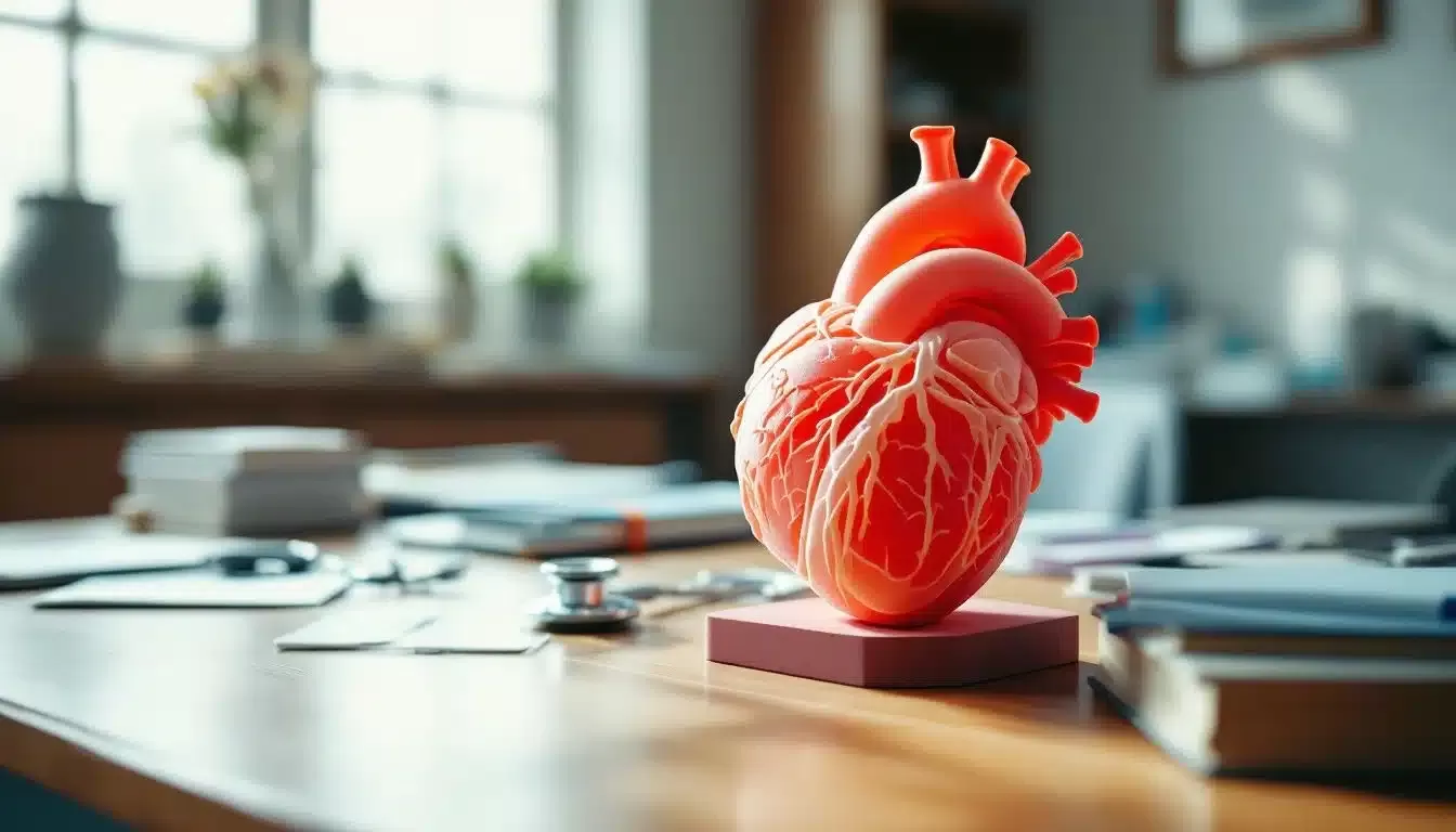
3D printing plays a pivotal part in making anatomical models. These lifelike replicas help medical students understand human anatomy more precisely and offer hands-on training.
The use of 3D printed anatomical models brings a new dimension to medical education. It allows for realistic, detailed study – enhancing learning experiences and improving surgical preparation.
Sure! The process begins with digital data from patient scans or generic anatomy databases… this information guides the printer as it builds up layers of material, crafting an accurate three-dimensional representation piece by piece.
While highly beneficial, some challenges exist , high-quality printers can be costly, and producing complex structures may require time… yet, many agree that the benefits outweigh these issues when considering enhanced understanding and improved patient outcomes.
Source: https://rapidmade.com/3d-printing-for-anatomical-models-transforming-medical-training/


)