Notifications

23 minutes, 45 seconds
-14 Views 0 Comments 0 Likes 0 Reviews
Female hormonal changes involved in gametogenesis, fertilization, and implantation. The follicle is known to be the functional unit of ovary and one female gamete; the oocyte is contained in each follicle.
What are the stages of oogenesis?
1. Oogonia, primary oocytes, secondary oocytes, and mature oocytes are the stages that the primordial germ cell passes through.
The primordial germ cells appear first in the dorsal wall of the endoderm of the yolk sac near the allantois as a cluster of 100 cells by 3-4 weeks of gestation. They then migrate from yolk sac to genital ridge through the hindgut and dorsal mesentery and colonize gonadal tissue by 7 weeks.
2. The oogonia
The primordial germ cells on reaching gonads are called as oogonia. The maximal oogonial content occurs when high mitotic activity undergoes fast mitotic multiplication, reaching 6-7 million by 16–20 weeks.
The primordial germ cells are seen here as white structures with black dots in it. Primordial germ cells are migrating towards the ridge where the gonad is. Look at during the first trimester. The first trimester where the oogonium where mitosis occurs with the oogonium and of course they are still deployed. When the oogonium still has 46 chromosomes, it undergoes its first meiosis. In the second trimester, the primary oocyte, which still has 46 chromosomes, goes through the leprotene, zygotene, pachytene, and diprotein stages of the first meiosis, but it still has 46 chromosomes. This secondary oocyte develops into a haploid oocyte at that point, marking the first instance in which these gametes have 23 chromosomes. The development of a mature oocyte and the ejection of the second polar body after the second meiosis are followed by the second meiotic division. The cumulus oocyte complex is produced as a result of ovarian stimulation.
The extracellular matrix caused the outer cumulus corona cells to split apart, and the corona cells next to the oocyte became less compact and began to radiate outward from the zona pellucida. At one o'clock, you may find the polar body one.
Oocyte loss with aging: Previously, there would be between 6 to 7 million oocytes between weeks 16 and 20 of pregnancy, but by birth, that number drops to 2 million, and by adolescence, it is around 40,000. However, in order to really ovulate, 400 oocytes are needed.
A recruited primordial follicle can grow and evolve into a specialized graphene follicle that has the ability to ovulate its egg into the oviduct through a process known as follicular genesis.
Phases of follicular growth:
· The primordial follicle, primary follicle
· The secondary or pre-antral follicle
· The antral and tertiary follicle, and
· finally pre-ovulatory follicle
A fully developed oocyte encircled by zona pellucida, six to nine layers of granulosa cells, a basal lamina, a theca interna, and a theca externa are its five unique structural components. When a hollow known as the antrum appears in the granulosa cells, it is the first sign that tertiary follicle growth has begun.
Pre-ovulatory follicle. A fully matured graphene follicle just prior to ovulation is about 20 millimeters.
Follicular fluid contains estrogens, FSH trace, amount of androgens, OMI, LI, inhibin, proteolytic enzymes and plasmid. Let's look at the two cell to gonadotrophin theory.
· The theca cell
· The granulosa cell.
Both of them have the LH receptors and the FSH receptors separated by the basement membrane.
What is required for the follicular recruitment?
· Initial recruitment, that is the primary part, the dominant primordial follicles are recruited into the growing follicle pool in a continuous manner.
· This increases in circulating FSH during each reproductive cycle recruit a cohort of antral follicles. Three theories are known.
One is theory of continuous recruitment, two single recruitment episodes and three follicular waves. This slide is very important because this will tell you that how do those follicles reach up to ovulation.
Please understand that the follicles that are going to be ovulating, say, for example, at the age of 40 years for the woman would be the oldest when compared to the ones that will actually ovulate at the age of 25. So therefore, those aged oocytes are not going to be as healthy as the oocytes that actually ovulate at the age of 25. So if you look at this slide, from the primordial to the primary follicle, the amount of time it takes to get recruited to the primary follicle can be any time and it's very difficult to guess, but from the primary to the secondary follicle, one will have to look at whether when the FSH receptors become active.
The initial recruitment from the secondary to the antral is something which could be anywhere between 120 days and from then on could be about 71 days. So from the antral follicle, which is roughly around 2 to 5 mm, the cyclic recruitment starts from antral, then it can go up. At every stage, you will have atretic follicles as well, which is apoptosis and then it moves forward where the selection and dominance occurs.
Once the selection occurs at the cyclic recruitment, it will be about 14 days, that's where all the gonadotrophins will act. But once that happens after 14 days, you will then have ovulation occurring. So this slide will just tell you that out of the many, many, many follicles that would have started for ovulation, very few will see the light of day coming towards ovulation.
The number of follicles that ovulate is determined by the length of FSH over the threshold, which we shall refer to as the FSH window. Increased FSH above the threshold is necessary for follicular recruitment. The second thing is, when there is a spontaneous cycle, the cycle of dominance allows for one follicle to claim the dominance and therefore does not allow for many follicles to actually move forward and ovulate and that's why in humans, you have singleton pregnancies more than multiple pregnancies because of the cycle of dominance. However, when you stimulate, you widen that threshold to a larger area and therefore on your right side, you will see a chart which shows multiple follicles growing only because you widen the window or the threshold so that many more follicles will be able to grow so that you got multiple follicles coming up.
Follicular selection is a process by which single dominant follicles is chosen from the recruited cohort or wave for preferential growth. Follicular divergence means that the time of selection, the growth profile of the dominant follicle begins to diverge as it continues to grow while subordinate follicles undergo atresia and this occurs when dominant follicle reaches around 10 millimeters on day 6 to day 9. The pre-ovulatory follicle reaches the status at about 16 to 29 millimeters in the late follicular phase. Estradiol production from the dominant follicle peaks and then results in an LH surge and followed by ovulation.
What are the reproductive hormones? The gonadotrophin-releasing hormone from the base and the LHRH hormone as well. It is a decapeptide. It is produced by the neurons with cell bodies in the arcuate nucleus and the gene encoding produces a 92-amino acid precursor protein which contains the JnRH decapeptide and GAP.
The pulsatility of GnRH has a short half-life of 2 to 4 minutes. Thus, continuous pulsatile secretion is necessary. The follicular phase frequent small amplitude pulses and the late follicular phase increase in both frequency and amplitude.
If you look in a spontaneous cycle to a conventional stimulated cycle, which means that you give exogenous gonadotrophins, in the first diagram on the left hand side, you will see the FSH level and you see that the threshold that you find, the window is quite narrow. So, you will see the arrows here that the follicles that are coming up, you will see only one arrow coming out and going further at the expense of all the other arrows which have already bent, which means that the window is small and there is only one arrow that has escaped that window threshold and then grown. Whereas, if you see a stimulated cycle, that window has enlarged and therefore a lot of follicles have been able to grow because there is extra stimulation because of exogenous administration of gonadotrophins and therefore multiple follicles are allowed to grow and this is what the stimulation does.
It allows for multiple follicular growth and takes out the single follicular dominance that happens in spontaneous cycles. The LH window means that once the follicles reach a critical size, then the LH surge gets activated. However, if the LH is suppressed, then the follicles tend to grow more and the cohort is also more.
So, therefore, a large number of cohort will be able to grow without allowing one of the follicles to get triggered so that the other follicles get luteinized, thereby allowing a good cohort of follicles to be taken for the pickup and many more follicles to be retrieved. This can happen if the LH is suppressed and that is why the GnRH is actually used. The AMH, that is the anti-Mullerian hormone, it's a member of the TGF family.
It's produced by the granulosa cells. AMH is produced by the growing antral follicles up to selection stage 4-6 mm reflect continuous non-cycling growth of small follicles. And this is a slide to show the interplay of all the four hormones that is required for the menstrual cycle, the estrogen, the LH, the FSH and the progesterone.
And you will see that there is positive and negative feedback here, all within the menstrual cycle and the red line is to show you the ovulation. And this has been described in my previous slides just to tell you that all these, if they are functioning in unison, you will be able to see that the ovulation occurs on the 14th day, the maximum peak of the estrogen would be just pre-ovulatory and then the progesterone peak would be somewhere around day 20 and of course, the ovulation occurs roughly around 36 hours before ovulation. So, 10 to 12 hours after the LH peak, the onset of LH surge, most reliable indicator of impeding ovulation occurs 36 hours prior to the follicular rupture and usually LH surge lasts around 48 to even 50 hours.
It takes two to tango, LH surge and FSH surge, resumption of meiosis oocyte, luteinization of granulosa cells, gives you progesterone, expansion of cumulus, prostaglandin synthesis, follicular rupture, LDL cholesterol, internalization, steroidogenesis and then into the corpus luteum. The FSH surge on the other hand, frees the oocyte from the follicular attachments, dispersion of cumulus freely floating in the enteral fluid, plasminogen converts to plasmin earlier by the proteolytic enzymes and the LH receptor on the granulosa cells is adequate for the luteal phase. The onset of LH surge is the most reliable indicator of impending ovulation occurring 34 to 36 hours prior to follicular rupture as shown in the diagram on the right.
This diagram, the physiology of migration of the sperms into the tube is seen in this particular diagram and the entire process is by thermotaxis, chemotaxis and rheotaxis. What is important is that the mobility of the sperms helps the sperms to move through the ova duct not only by itself but also by the peristalsis of the tube. It takes about five days to seven days for the fertilization to occur after the release of the ovum and as it reaches the ampulla where the fertilization occurs, the sperm meets the ovum and there is where this fertilization occurs and if you look at the embryo nutrition here, the ova ductal epithelial cells, there you will find glycogen, the amylases and of course and you'll find oxidative metabolism, the pyruvate and lactate, the fertilized embryo which is here, the immature mitochondria, you'll see that the glucose and the pyruvate at the cleavage stage embryo and the embryo development occurs.
You'll also see all the other factors that have been depicted here and as you reach the blastocyst stage, you will see the glycolytic metabolism which you'll see with mature elongated mitochondria and of course you will see the glucose, the oxygen and of course release of ATP. Protection of embryo in the tube, you'll see that as you go through embryo in the tube, you will see the oxidative stress there, you will also find the heat stress there and the immune response, the antimicrobial peptides and excess protease activity. So, there is certain responses that you will see and the immune response also to protect the embryo.
How does it get protected?
· One, it protects against the oxidative and heat stress with oestrogen-mediated embryo protection against the immune system and the embryotropic factors like growth factors for the cleavage and development.
· In the transport, fluid secretion, fluid flow and ciliary movement, the nutrients are pyruvate, glucose, glycoproteins and lactate and the epigenetic regulation like the DNA methylation, histone modification. So, all these things help to protect the embryo but through the oviduct.
The time of the fertilization to the time it reaches to the endometrial cavity would be anywhere between six to eight days when the implantation occurs. So, the implantation is about a week after fertilization. The embryo wastage is something that we need to understand, the iceberg in the human reproduction. This is the rate limiting step and a bottleneck of reproduction. Success rates in art have never crossed over 40 percent and of course, the live birth rate is about 30 percent.
At the pre-implantation loss itself is 30 percent and post-implantation another 30 percent, clinical pregnancy loss about 10 to 15 percent. So, therefore, we have a loss which is something that we have to be careful about. So, we are not as great a reproducer as the mammalians or as cats and dogs because they have litters. So, our loss is much higher and we are actually poor reproducers. So, even with euploid embryos, our success rate is never beyond 70 percent. So, who are the contributors for a successful implantation? A competent embryo that is a blastocyst, an embryo endometrial synchrony, receptive endometrium, autocrine, paracrine, endocrine factors, embryo maternal signalling and finally, a successful implantation.
What are the morphological steps of implantation?
· Apposition
· Adhesion
· Invasion
The embryo will first have to oppose the lining, then it will have to stick to the lining and then it will have to invade and that is when the trophoblast takes over. So, continuous process from conception to about 22 weeks of gestation is what happens with these three processes.
However, as complex as it may seem and as simple as it's been explained, nevertheless this blog has covered it all. The more we know, the more we have to know – so never stop learning.
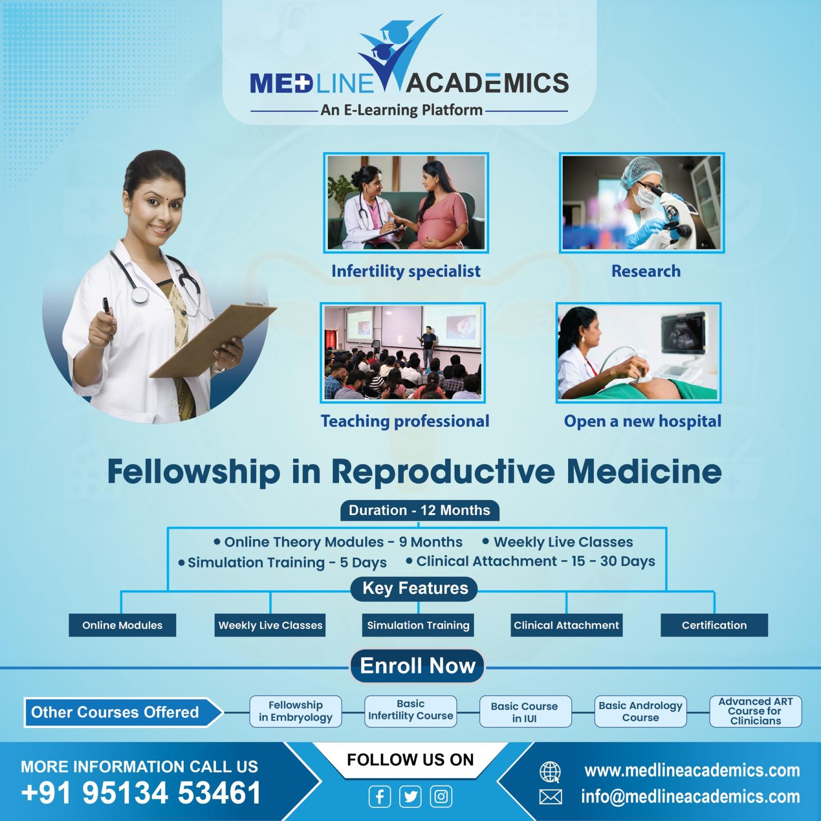
A comprehensive Infertility and IVF Fellowship Courses in India is offered by Medline Academics for medical professionals worldwide. This program combines self-paced online learning with interactive webinars led by renowned experts, making it ideal for practicing doctors looking to expand their expertise in infertility. The curriculum includes structured modules, real-life case studies, and a graduation certificate upon completion, ensuring continuous professional development. Medline Academics' fellowship provides healthcare practitioners with the latest knowledge and skills to excel in the field of reproductive medicine, while emphasizing accessibility and quality.
Additionally, Medline Academics offers a highly accessible online fellowship in reproductive medicine in India for healthcare professionals worldwide. The fellowship offers a rich learning experience through digital modules, expert-led discussions, and hands-on clinical insights that seamlessly integrate with a busy medical practice. A participant not only gains theoretical knowledge, but also practical skills that can be used in patient care immediately. Through innovative, web-based platforms that support lifelong learning, Medline Academics advances reproductive healthcare education.
A leading IVF Hospital in Bangalore, Dr. Kamini Rao Hospitals is well-known for providing top-notch fertility treatments based on cutting-edge medical technology and a kind, patient-centered philosophy. The facility was created by the renowned Dr. Kamini Rao with the goal of providing couples with outstanding reproductive healthcare that respects their individuality and mental wellness as they prepare to become parents. IUI, IVF, ICSI, and fertility preservation are just a few of the fertility procedures offered by the facility, which are all customized to each patient's specific need. The facility guarantees the greatest results and high success rates because to its cutting-edge assisted reproductive technologies. A committed group of fertility doctors, embryologists, and counselors who provide individualized support and knowledgeable direction throughout the course of therapy form the foundation of its care approach. Dr. Kamini Rao Hospitals, one of the most reputable and prosperous fertility clinics in India, maintains its standing as a top IVF facility in BLR where compassionate treatment and high-quality medicine coexist.
Fellowship in Reproductive Medicine IVF Hospital in Bangalore IVF Center in Bangalore Dr Kamini Rao

